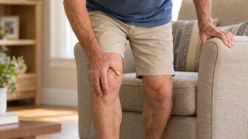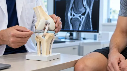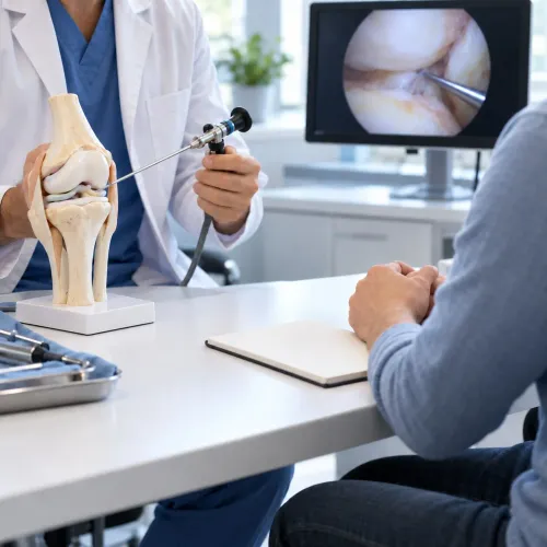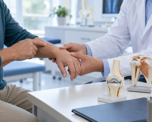
The same knee, hip, or shoulder pain can come from a focal cartilage defect, unstable ligament, meniscal tear, abnormal alignment, early arthritis, or a problem outside the joint. By the end, you will know how clinicians test whether your natural joint is salvageable, compare preservation with replacement or non-operative care, and judge the recovery commitment before choosing treatment.
Key takeaways
- Preservation works best before widespread cartilage loss and advanced arthritis.
- Treatment may combine cartilage repair, alignment correction, ligament work, or meniscus surgery.
- Replacement becomes more suitable when pain and joint damage are widespread.
- Ask about recovery milestones, follow-up imaging, alternatives, and a second opinion.
What does joint preservation surgery include, and what problem is it meant to solve?
Joint preservation surgery is a treatment category, not one operation. It aims to keep your native joint functioning and postpone or avoid partial or total replacement.
Depending on the problem, it may combine cartilage restoration for a focal defect, meniscus repair, ligament reconstruction for instability, an osteotomy to correct abnormal loading, core decompression for selected bone damage, or hip preservation procedures such as arthroscopy.
| Problem | Preservation approach | Planning question |
|---|---|---|
| Focal cartilage defect | Cartilage restoration | Is the defect well defined, with limited surrounding arthritis? |
| Meniscus or ligament injury | Meniscus repair or ligament reconstruction | Is mechanical damage causing pain, catching, or instability? |
| Bow-legged or knock-kneed loading | Unloading osteotomy | Will alignment continue damaging the repaired area? |
| Hip impingement or early damage | Hip preservation procedure | Is the pain mechanical rather than established diffuse arthritis? |
| Bone circulation problem | Core decompression | Is the bone still suitable for preserving the joint surface? |
The operation must target the true pain source, not simply an abnormality seen on a scan. A cartilage defect can be incidental; pain may instead come from synovitis, a bone-marrow lesion, meniscal pathology, tendinopathy, referred spine pain, or generalized osteoarthritis. A cartilage procedure cannot compensate for untreated malalignment or instability.
That distinction matters because technically successful surgery still disappoints when the diagnosis is wrong. A focal defect differs from widespread cartilage loss, marked joint-space narrowing, inflammatory arthritis, or uncontrolled loading, where preservation is less likely to solve the problem.
How do surgeons decide whether a joint is still salvageable?
A joint is considered salvageable when pain comes from a treatable, local problem and enough healthy joint remains to support repair or realignment. Patient selection weighs symptoms, examination findings, activity goals, general health, and your ability to complete a prolonged rehabilitation program before choosing preservation over partial or total joint replacement.
Surgeons look for:
- A focal cartilage or meniscus defect rather than widespread cartilage loss
- Preserved joint space and usable cartilage elsewhere in the joint
- Correctable limb alignment and acceptable bone quality
- Stable or reconstructable ligaments
- A body weight, smoking status, and medical condition compatible with healing
- Symptoms that match the suspected structural problem
- A realistic willingness to follow restrictions, physiotherapy, and protected weight-bearing
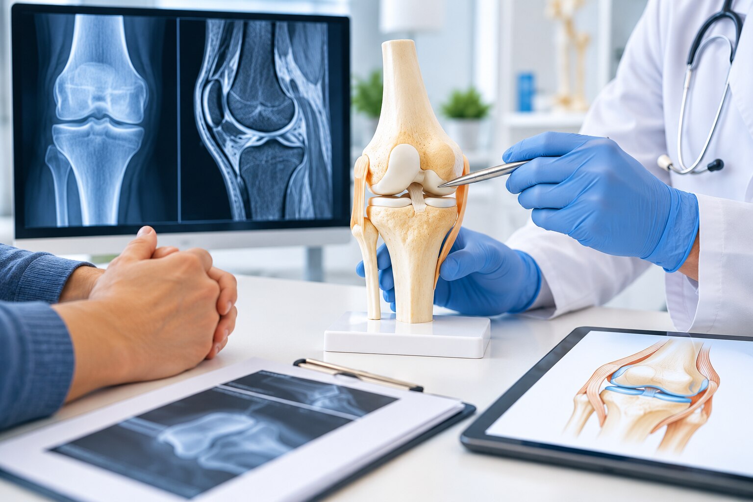
The diagnostic work-up begins with a physical examination, gait assessment, and review of previous treatment. Weight-bearing X-rays show joint-space narrowing, alignment, bone spurs, and arthritis under load; an MRI assesses cartilage, menisci, ligaments, labrum, and bone marrow. A CT scan adds detail when bone shape, impingement, or alignment needs precise measurement.
Imaging alone cannot show how much pain you feel, because structural abnormalities can exist without symptoms.
Preservation becomes less suitable when arthritis is diffuse, joint-space loss is substantial, alignment cannot be corrected, bone quality is poor, or instability cannot be controlled. Increasing age does not automatically exclude treatment, but advanced arthritis and limited rehabilitation capacity make lasting benefit less likely than replacement.
How does the treatment plan change for cartilage, alignment, ligament, meniscus, hip, and early arthritis problems?
The diagnosis determines which structure must be corrected; treating pain alone will not preserve a joint. A conservative trial is adequate when you complete the prescribed physiotherapy, follow activity or weight-management advice, use suitable medicines or injections when offered, and return for reassessment with documented function and pain.
Bracing applies when it unloads an affected compartment or supports instability, not as a universal treatment.
| Problem | Preservation strategy | What an adequate non-operative trial means |
|---|---|---|
| Focal cartilage defect | A small, contained defect may suit microfracture; larger or deeper defects may need an osteochondral graft. | Physiotherapy, impact reduction, prescribed medicine, and weight management when relevant; persistent focal symptoms support further planning. |
| Meniscus tear | A repair preserves shock absorption; removing damaged tissue is less protective. | Rehabilitation and activity modification for a stable tear; locking, displacement, or persistent symptoms can change the decision. |
| Malalignment | An unloading osteotomy redirects force away from worn cartilage. | Strengthening, weight reduction, brace use, and activity changes; these do not correct bone alignment. |
| Ligament instability | Ligament reconstruction may be combined with cartilage or meniscal treatment. | Structured physiotherapy and a brace for suitable stable injuries; repeated giving-way favours reconstruction. |
| Hip impingement or labral damage | Hip arthroscopy can reshape the impingement lesion and repair or preserve the labrum. | Hip-specific physiotherapy, movement changes, medicines, and selected injections; established joint-space loss makes arthroscopy less suitable. |
| Early arthritis | Combine strengthening, weight management, low-impact activity, medicines, injections, or a brace before considering preservation surgery. | Reassess after an adherent, diagnosis-specific programme; diffuse cartilage loss is not a focal cartilage problem. |
A cartilage operation cannot overcome untreated instability or abnormal loading. The plan must address every mechanical driver at the same time.
When is preservation a poor choice, and how does it compare with replacement?
Preservation is a poor choice when pain comes from widespread bone-on-bone arthritis, severe deformity, uncontrolled malalignment, inflammatory arthritis, or a joint that cannot heal reliably. Operating on the wrong pain source can leave symptoms unchanged while adding stiffness, infection, blood clots, nonunion, or recurrent instability.
| Option | Pain relief | Recovery and activity | Future surgery |
|---|---|---|---|
| Non-operative care | Often reduces symptoms without removing structural damage | No surgical recovery; activity changes, physiotherapy, medicines, injections, or bracing may be needed | Arthritis may progress; replacement remains possible |
| Preservation surgery | Best for a specific, treatable defect; relief is less predictable with diffuse arthritis | Usually the longest recovery; crutches, protected weight-bearing, or a brace may last weeks, with running and pivoting delayed for months | May delay replacement, but failure or arthritis progression can lead to later surgery |
| Partial joint replacement | Usually reliable relief when disease is confined to one compartment and ligaments remain suitable | Faster recovery than many preservation procedures; some impact and kneeling limits may remain | The implant can wear or the remaining joint can deteriorate, requiring revision or total replacement |
| Total joint replacement | Most predictable option for advanced arthritis and severe pain | Recovery is structured and progressive; high-impact sport is commonly discouraged | Implant loosening, wear, infection, or fracture can require revision |
A focal cartilage defect with good alignment may justify preservation, but a severely damaged joint often gains more from replacement. Choose based on the pain generator, imaging, healing capacity, and the recovery restrictions you can realistically follow—not on age alone.
What recovery, follow-up, and second-opinion questions should you plan for?
Expect a longer, more restrictive recovery than the operation name suggests: your hospital stay may be same-day or overnight, while crutches, a brace, and protected weight-bearing can continue for several weeks.
1. Ask, “How long will I use crutches or a brace, and when can I fully bear weight, bend the joint, climb stairs, and drive?” Driving requires safe control of the vehicle and no sedating pain medicine; your insurer may impose additional rules.
2. Ask for written work and rehabilitation milestones, including when desk work, standing, lifting, and physical work are realistic. Physiotherapy may begin early, but running, jumping, and pivoting are delayed until strength, range of motion, balance, and functional tests are satisfactory.
3. Ask, “What is your return to sport test, and what restrictions remain?” Return to sport can take many months, and elapsed time alone does not prove readiness.
4. Ask which warning signs require urgent review: fever, wound drainage, calf swelling, chest pain, sudden breathlessness, worsening stiffness, or recurrent instability. Infection, blood clots, incomplete healing, and arthritis progression can lead to further treatment.
5. Ask how failure would be managed, including whether partial or total replacement could later be required, and how that outcome compares with non-operative care. Ask for the surgeon’s experience with your exact procedure, expected success criteria, rehabilitation burden, and alternatives.
UR Orthopaedic Clinic can help coordinate assessment when you want these questions answered alongside examination and imaging. Take your scans and reports for a second opinion before consenting.
Frequently asked questions
What does joint preservation surgery include?
It includes procedures that protect or restore the native joint, such as cartilage repair, osteotomy for alignment, ligament reconstruction, meniscus treatment, and selected hip-preservation operations.
How do surgeons decide whether a joint is salvageable?
They assess your symptoms, activity goals, physical examination, X-rays, MRI findings, alignment, stability, cartilage damage, and the extent of arthritis.
How does treatment planning differ between cartilage, alignment, ligament, and meniscus problems?
Cartilage defects may need repair or restoration; malalignment may require bone correction; ligament injuries may need reconstruction; and meniscus treatment depends on tear location, tissue quality, and joint damage.
When is joint preservation a poor choice?
It becomes a poor choice when arthritis affects most of the joint, cartilage loss is extensive, deformity is severe, or symptoms cannot be addressed without replacing the joint.
What should you ask about recovery and a second opinion?
Ask about weight-bearing limits, brace or crutch use, physiotherapy, return-to-work and sport timelines, follow-up imaging, complications, alternatives, and the surgeon’s reason for recommending preservation.